Even before I met with my doctor, I had noticed some symptoms that I was worried might be indicative of a problem. I had been having trouble with stumbling and I had really been trying to get back in shape and doing power walking during the spring. So the stumbling worried me. After I saw my NS in June, I started feeling weird around July 4 when we went walking downtown with friends. I was finding it really uncomfortable to be around the crowds. I just wanted to go home. Also didn’t like all the noise from the crowds (there was a July 4 festival with music, lots of people, food, etc.)
By August, I was definitely having headaches and dizziness again. I called my doctor’s office. Wasn’t sure which doctor to ask for since I’d had one before but had been passed on to the head doc. Called his assistant who was extremely rude to me and said she would have him look at the MRI but she couldn’t talk to me because she was dealing with someone else.
I later heard from my other doctor’s Physician Assistant…
August 8: Kristen, Dr. K’s PA
”Dr. B sees nothing that needs surgical treatment. Adhesions (scar tissue). They are higher up and wouldn’t be causing Chiari symptoms. Occasionally adhesions are compressing. Mine aren’t compressing- they are just there. B is expert guy re: Chiari so will be seeing him in future, not K. No need to do Cine MRI followup in 6 months.”
She told me to go see a Neurologist.
So I’m thinking- WHAT? But I called my Neurologist. Who I hadn’t seen in a year. Amazingly I managed to get in to see her August 14th- so there must have been a cancellation.
FOUND THIS INFORMATION ON THE INTERNET: Individuals require periodic follow up after surgical treatment for a Chiari malformation. Symptoms may recur after a successful surgery, usually within the first two years. Most likely, this is due to the development of scar tissue or an opening around the duraplasty covering the brain.
August 14: Met with Neurologist along with my hubby, who keeps notes on things and keep me from having a total meltdown. Dr. C looked at my photo that I had on my phone (below) of the MRI and said “That is a CYST!” 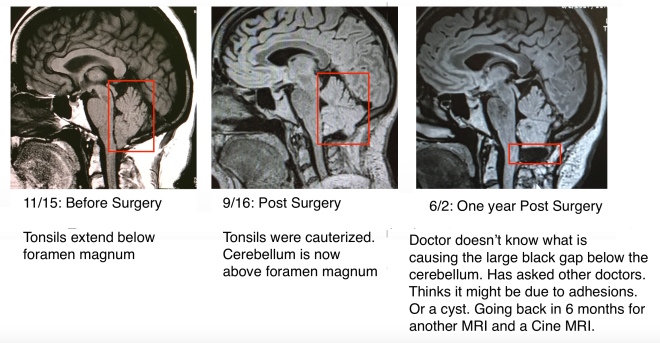
She referred me to a MRI and CINE MRI. I was very shaky at the appointment and my hands were shaking. Could have been low blood sugar though. But was dizzy and unbalanced.
August 15th I had one MRI. Results of that came back and didn’t see anything significant. BUT THEY WERE COMPARED TO AN MRI I HAD A YEAR AGO and NOT the one I HAD on JUNE 2 as they were done at different hospitals. This is really a problem as the docs can’t seem to get all the pertinent information together so everything can be accurately compared!!
I was due to have a Cisternogram on Friday the 18th, but the hospital called and cancelled it and said that it was the wrong test. I needed a CINE MRI. At least they managed to let me know it was the wrong one before I went there like last year where I was all prepped for the test and they said it was the wrong test. I DON’t need a Nuclear Cisternogram test where they inject dye in your spinal cord to check for leaks. I need a CINE MRI to check CSF flow in the brain. I said this to the person from the hospital and to my Doc’s Asst. HOW COME I KNOW MORE ABOUT THIS FREAKING TEST THAN THEY DO??
Anyway, managed to get the CINE MRI on August 28th. ……
It said…..
“There is a normal CSF slow signal anterior to spinal cord at the level foramen magnum. Posteriorly, however, there is near absence of signal posterior to the cord and within the patulous CSF space interior to the cerebellum. There is markedly diminished signal in the fourth ventricle or cerebral aqueduct.
In the lower level within the upper cervical spine at the level of C1-2, there is a normal signal anterior to the spinal cord, as well as mildly reduced signal posterior and lateral to the cord (see axial view).
IMPRESSION:
1. Stable postsurgical changes of prior Chiari decompression with a suspected loculated CSF space inferior to the cerebellum with synechiae. While there is no clear anatomical obstruction at the C1 laminectomy site, this excluded posterior fluid collection likelyresults in reduced CSF flow velocity and causing apparent absent CSF flow signal posterior to the cord at the foramen magnum.
2. No evidence of CSF obstruction anterior to the cord at the foramen magnum.
I called my Dr’s office once the results were in and asked what they recommended. Always antsy waiting for results. Dr. was referring me to see a NS at a different hospital and not at Goodman Campbell.
So I called up Goodman Campbell and asked to speak to the office manager and told her how Dr. B. had BLOWN ME OFF and how I got the CINE MRI which said I have posterior CSF flow blockage!!
I then got a call from DR. K. a day or so later and he started off saying the same old line they gave me before…. I said well I had the CINE MRI that YOU DIDN’T want to ORDER UP and it says BLOCKAGE OF CSF FLOW!!! So he said he see if I could have a consult with another NS there. …..